Advanced Heart & Vascular Imaging Center
Comprehensive Cardiovascular Imaging.
Conveniently located in Philadelphia.
ReVaMP Heart & Vascular Imaging Center is expanding access to advanced cardiovascular imaging with a comprehensive range of diagnostic services available onsite at our Philadelphia imaging center.
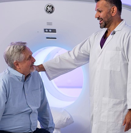
Comprehensive Cardiovascular Imaging Under One Roof
From coronary artery imaging and calcium scoring to PET-CT, SPECT, ultrasound and echocardiography, our advanced imaging services help physicians evaluate heart and vascular health with greater detail and efficiency—all in one convenient location.
Our Onsite Cardiovascular Imaging Services
PET-CT combines positron emission tomography (PET) with computed tomography (CT) to provide functional and anatomical information. Depending on the clinical indication, PET imaging can help physicians evaluate blood flow and heart function and identify areas that may require further evaluation.
Single-photon emission computed tomography (SPECT) is a nuclear imaging test that can help evaluate blood flow to the heart muscle and identify areas of reduced blood flow, particularly during cardiac stress testing.
Coronary CT angiography (CCTA) is a noninvasive imaging test that creates detailed images of the coronary arteries. It can help physicians evaluate the presence of plaque and narrowing in the coronary arteries and assess cardiovascular risk.
A coronary calcium score uses a CT scan to detect and measure calcium deposits in the coronary arteries. The results can help physicians assess the presence of coronary atherosclerosis and better understand an individual's cardiovascular risk.
Peripheral CT angiography provides detailed images of blood vessels and surrounding structures outside of the heart. It can assist physicians in evaluating the peripheral vascular system and identifying potential areas of narrowing or other abnormalities.
An echocardiogram uses ultrasound waves to create moving images of the heart. It can provide important information about heart structure, heart function, valves and blood flow.
Ultrasound uses sound waves to create images of structures within the body without radiation. Vascular and other ultrasound examinations can help physicians evaluate blood vessels and identify potential abnormalities.

Advanced Imaging. Without the Long Waits.
When cardiovascular imaging is needed, convenience matters. ReVaMP brings multiple diagnostic imaging services together at one Philadelphia location, making it easier for patients and referring providers to access the imaging they need.
Why Choose ReVaMP?
Whether imaging is being used to investigate symptoms, evaluate cardiovascular risk, monitor an existing condition, or support a treatment plan, ReVaMP provides advanced diagnostic imaging to give you a more complete picture of your heart and vascular health. Our cardiologists use these insights, along with your medical history and individual risk factors, to better understand your cardiovascular health and develop a personalized approach to your care.
Comprehensive
Access multiple cardiovascular imaging services at one convenient location.
Advanced
Utilize sophisticated imaging technologies designed to provide detailed information about the heart and vascular system.
Convenient
Located in Philadelphia for easier access to advanced cardiovascular diagnostics.
Imaging and Expertise, Together
ReVaMP cardiologists interpret your diagnostic imaging, evaluate your individual cardiovascular risk factors, and develop a personalized treatment plan designed around your cardiovascular health.
ReVaMP Heart & Vascular Institute Imaging Center
Philadelphia
510 N Broad St Suite 202
Philadelphia, PA 19130
Phone
(267) 541-4774
Fax
(833) 455-8188
Hours of Operation
Wednesday: 8:00 AM – 5:00 PM
Thursday: 8:00 AM – 5:00 PM
Friday: 8:00 AM – 5:00 PM
Five Greentree Centre, 525 NJ-73, Marlton, NJ 08053, USA
2418 E York St, Philadelphia, PA 19125, USA
170 N Henderson Rd, King of Prussia, PA 19406, USA
727 Welsh Road, Huntingdon Valley, PA, USA